🧲 New MRI Technique to Speed Up Multi-Contrast MRI Scans and Advance Early Alzheimer’s Biomarker Discovery
A new publication from PREDICTOM introduces a promising method to dramatically accelerate multi-contrast brain MRI scans, without sacrificing image quality. The technique could pave the way for earlier and more accessible diagnosis of neurodegenerative diseases like Alzheimer’s disease.
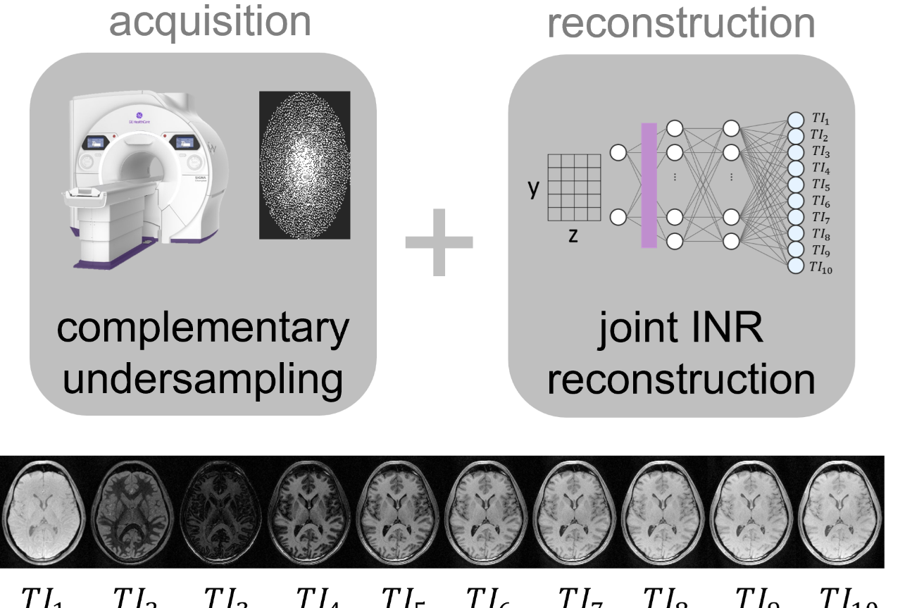
Overview of the proposed method and resulting multi-contrast MRI images for the MPnRAGE sequence
This new publication is presented at the International Conference on Medical Image Computing and Computer-Assisted Intervention (MICCAI) 2025 by Natascha Niessen, PhD student at the Technical University of Munich, the work was developed with her team at GE HealthCare. It explores how multi-contrast MRI sequences, which provides rich information about brain tissue, can be acquired faster using advanced undersampling and a novel deep learning reconstruction approach called Implicit Neural Representation (INR). Preliminary work to this study was also presented at the Conference of the International Society for Magnetic Resonance in Medicine (ISMRM) earlier this year.
What is Multi-Contrast MRI?
Multi-contrast MRI captures several images of the same brain using different MRI contrast settings. These images can be used to estimate quantitative tissue properties, offering the potential to detect subtle changes in brain microstructure. However, the scan time of multi-contrast MRI can be very long, which can lead to motion artifacts and limit clinical use.
How Can MRI Be Accelerated?
One way to speed up MRI scans is undersampling, which means collecting fewer data points. However, undersampling can lead to blurry or incomplete images unless advanced reconstruction techniques are used. In multi-contrast MRI, the same brain is scanned multiple times with different settings, so the different images differ in contrast but share anatomical information.
This opens up the possibility of complementary undersampling, where different contrasts are undersampled in a coordinated way. The INR deep learning architecture takes advantage of this shared anatomical structure to jointly reconstruct all contrast images, resulting in high-quality scans even with much less data.
The INR network is self-supervised and scan-specific, meaning that it can reconstruct the undersampled raw MRI data without the need for any training data. This is particularly suitable for multi-contrast MRI where large datasets are scarce.
“We’re able to reconstruct high-quality images from highly undersampled data by leveraging shared anatomical information across contrasts. This means shorter scan times and potentially better access to multi-contrast MRI in clinical settings,” says lead author Natascha Niessen, PhD student at GE HealthCare and Technical University of Munich.
Making Multi-Contrast MRI Feasible for Clinical Studies
By enabling faster acquisition of multi-contrast MRI scans, this technique allows more patients to be scanned with this kind of advanced imaging protocol. This supports large-scale studies and facilitates the discovery of novel biomarkers for neurodegenerative diseases such as Alzheimer’s. To translate this into clinical research, the team is integrating complementary undersampling into the multi-contrast MPnRAGE sequence, which will be part of the advanced MRI protocol in the PREDICTOM study.
Natascha Niessen


